In 2005, I experienced an odd visual problem. Yes, 2005 was a long time ago, but I still remember it well. I was on my first teaching prac at the time. One evening, when I was trying to write up my day's notes and prepare for the next day, I noticed a strange spot in my vision in my left eye. There seemed to be a small ‘bubble’ distorting whatever I was looking at behind that spot. Something a little bit like this…
Ok, it's hard to draw. That's not quite right but it gives you some idea. Anyway, I put it down to my stress and tiredness (prac teaching is very hard work) and went to bed. The next morning it was still there, but it seemed to gradually fade over the following days.
Then a few years later, I noticed another interesting effect in the same region of vision in my left eye. A kind of wrong-colours and shimmering effect. You can read about this in a blog post I wrote in 2009, including another attempt to show you what I saw with an edited image. My ophthalmologist at the time (not the one I saw recently) suggested it was some kind of retinal pigment epithelium (RPE) defect, but wasn't sure what might have caused it.
Over the following few years, I noticed another patch appearing in my right eye, closer to the centre. The visual appearance changed gradually. Both eyes now have several spots with problems, some bigger than others. Let me try another vision-map like the one in this progress report blog post from 2011.
Ok, that's not perfect either, but again it should give you some idea. Notice that I've drawn this as I see it, which is actually opposite to how things are normally done in the medical field. Normally these things are done ‘as the doctor sees it’ looking at you.
The shimmering has now reduced but occasionally reappears; when it does, it usually lasts a few days. Colours are not right in these areas. Lines and shapes are distorted through them. I also see some reduced-contrast flare if there's direct sunlight falling across my eyes (eg. if the sun is rising or setting to the side).
Anyway, when I went back to the ophthalmologist recently, he made a suggestion about what might have been going on. He said it could be something called choroidal serous retinopathy (CSR). I looked this up and I think it does describe my symptoms. (Actually, most of what you'll find with a google search is central serous chorioretinopathy, which is basically the same thing only near the fovea, which is the part of the retina behind the centre of where you look—the + signs in my diagram above.)
<
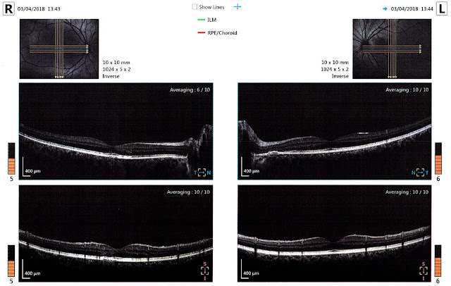
What evidence is there that this might be my problem? I'm glad you asked. Here's part of the OCT scan taken of my retinas at my ophthalmology appointment in April:
Ok, so maybe you don't know how to read one of these. I already had some idea since doing some research after my first OCT years ago, but reading this page helped enormously—go read it if you're interested in more detail than I talk about here.
- These images are ‘standard’ medical imaging, so my left eye is on the right, and my right eye is on the left.
- The small thumbnails at the top are retinal photos with lines indicating how the machine scanned my eyes. The middle row is a horizontal scan (cyan arrow in thumbnail). The lower row is a vertical scan (magenta arrow in thumbnail).
- N/T in the middle row is Nose/Temple. Looking left to right, the right eye scan goes from temple (side of face) to nose (middle), then the left eye scan from nose (middle) to temple (side). The centre of the page shows the head of the optic nerve, corresponding to the blind spot. When you look for it in your vision, the blind spot appears towards the outside from the centre, but remember the image on your retina is inverted; the nerve is actually closer to your nose.
- Similarly, S/I in the bottom row is Superior/Inferior, ie. top/bottom. That is, the top of the retina is on the left in the scan, and the bottom on the right. Again, remember that this is the opposite of what you see: the top of your retina sees the bottom of your vision, and vice versa.
- Finally, the dip in the middle of each scan is the fovea. The centre of your vision has much higher resolution (actually ‘acuity’), meaning many more light detecting cells. The shape of the retina here is to make room for this.
The highlighted sections (and maybe also the bit with a dimmer highlight) correspond approximately with where I see visual issues in my left eye, out from the centre towards the blind spot. The left-most section highlighted here certainly looks to me (a non-expert) like the OCT examples for RPE CSR from this page I linked to above.
And here's my right eye scans near the fovea:
Again, the highlighted sections correspond to my visual issues in my right eye, just outside and below the centre (so inside and above the fovea on my retina).
Whew! That was pretty technical.
<
So what does this all mean? Well, interestingly, two potential causes stood out for me from my research: stress and steroids. (Actually, they're correlated with CSR, but not necessarily causal.)
Stress. My life is quite stressful. Teaching is stressful (and prac teaching, when I first noticed a problem in my vision, even more so). I'm also a middle leader at work (head of faculty). And I'm studying part time. Maybe I need to reduce my stress. But how—what should I change?
Steroids. I've been using corticosteroids for asthma since I was a teenager. I use corticosteroids to reduce my rhinitis. I also use topical corticosteroids at times to treat eczema and a variety of related skin problems. Probably not something I can easily avoid.


